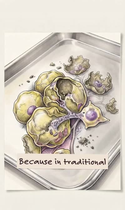
The concept the slide states first
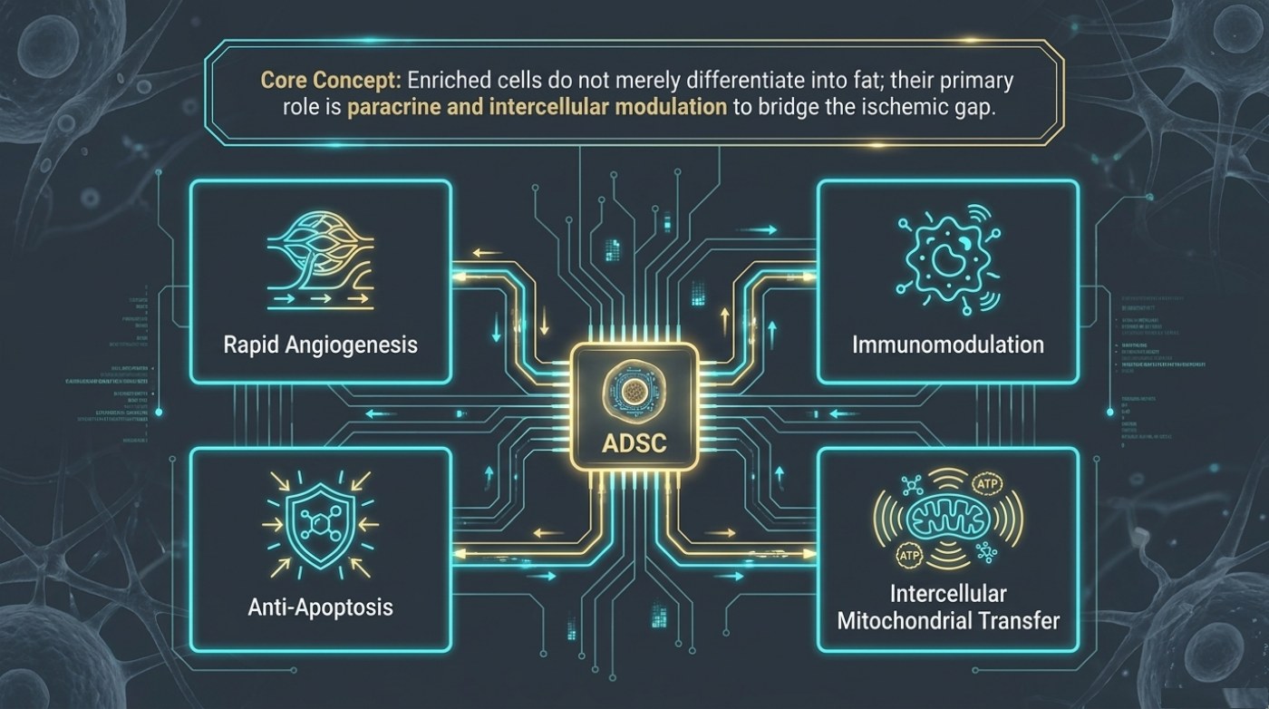
The banner above the diagram in deck drop2_3 gives the concept before the four boxes. Enriched cells do not merely differentiate into fat, the slide says; their primary role is paracrine and intercellular modulation to bridge the ischemic gap. That sentence answers a common assumption. The added cells are not described as turning into new fat cells to replace what was lost. They are described as signalling: releasing factors that act on nearby tissue, and exchanging material directly with neighbouring cells.
The short version
- A fat graft lands with no capillary network, so rapid angiogenesis is placed first among the four support mechanisms.
- The briefing describes enriched cells acting through paracrine signalling and intercellular modulation rather than becoming replacement fat.
- Vessel growth shortens the one-to-three-day interval the tissue can tolerate without perfusion rather than boosting final volume directly.
- The material names VEGF and FGF2 among the pro-angiogenic factors and a shift of macrophages toward the anti-inflammatory M2 phenotype.
- Angiogenesis carries no retention figure; the retention gains above 60% belong to the preparation slide and its named products.
Paracrine means the effect is local and chemical, with a cell releasing factors that act on the cells around it rather than on itself. Intercellular modulation means the exchange happens between cells in contact. Both are mechanisms of influence rather than of replacement, and the four labelled boxes around the central node describe the forms that influence takes.
| Common assumption | What the briefing describes instead |
|---|---|
| The added cells become new fat cells | Their primary role is paracrine and intercellular modulation |
| The cells replace the graft’s blood supply | They prompt vessel growth so the graft can establish its own perfusion |
| The support is a one-off at the moment of transfer | The support is aimed at a specific window of roughly one to three days |
| Success means the added cells survived | Success means the graft survived |
Why angiogenesis is placed first
Rapid angiogenesis sits at the upper left of the diagram, and the ordering is not decorative. A transplanted graft is avascular at the moment it lands. Whatever else is happening in the tissue — inflammation, cell stress, the reshaping of the scaffold around the graft — none of it can be resolved without perfusion. Until vessels reach the tissue, every other mechanism is working against a clock.
| Mechanism, as labelled on the slide | What it acts on | Why it sits where it does |
|---|---|---|
| Rapid angiogenesis | Vessel growth into and around the graft | First, because the graft has no capillary network when it lands |
| Immunomodulation | The local immune response in the graft’s first days | The early phase is an inflammatory event |
| Anti-apoptosis | Stressed cells at risk of dying | Viability is the quantity the resorption curve tracks |
| Intercellular mitochondrial transfer | Energy-carrying components passed between cells | Support for cells that cannot generate enough energy themselves |
The slide’s fourth box carries the label intercellular mitochondrial transfer, and the diagram attaches ATP to the mitochondria it shows. That is the most direct of the four mechanisms: rather than a signal released into the space between cells, functioning components move from one cell to a stressed neighbour. The site’s published guide to fat survival describes the same idea in patient-facing terms, naming mitochondrial transfer between cells among the mechanisms the source material gives.
The word rapid is the argument
What matters about angiogenesis here is not that it happens but when. The graft’s problem is not the absence of vessels in principle; it is the length of time before they arrive. The briefing places the tissue’s tolerant interval at roughly one to three days, which is the same window the resorption slide draws its steepest decline through and the same gap the paradigm slide describes cellular support as bridging. Faster vessel growth does not improve a graft’s final volume directly. It shortens the period during which volume is being lost.
That is why the mechanism is better described as enabling than as producing. Angiogenesis restores the conditions under which the rest of the graft can behave normally. The published guide names the signalling the source material describes behind two of these boxes: pro-angiogenic growth-factor secretion, with VEGF and FGF2 among the factors named, and a shift of macrophages toward the anti-inflammatory M2 phenotype. It also records the outcome the source material attributes to culture-expanded cells — retention gains above 60% — as a figure from that material rather than a measured constant.
Where this sits in the wider argument
This slide is the last piece of the briefing’s biological case. The resorption slide establishes that conventional grafting loses volume on a schedule. The paradigm slide names the approach of adding the patient’s own cells. The preparation slide separates the two products that approach can mean. This slide explains what those cells are supposed to do while the graft has no blood supply. Read together, the four make one claim: the interval without perfusion is the variable, and the biology is aimed at it.
Four boxes, one target
The diagram places one node at its centre and four labelled boxes around it, and the connecting lines run in both directions rather than as a chain. The slide does not state an order in which the mechanisms fire, and it does not rank them. What it does state is their shared aim: the interval before perfusion returns. Angiogenesis is the one that ends the interval, and the other three describe what is being done for the tissue while the interval lasts — acting on the graft and its neighbours rather than on the recipient site as a whole.
For a reader, the practical consequence is a question rather than a mechanism to memorise. Material that names a single pathway and attaches a volume figure to it is compressing four labels into one claim this slide does not make. In the briefing the mechanisms belong to the biology and the retention figures belong to named products, which is why they are described on separate slides. Keeping them apart is not pedantry; it is the difference between reading a result and reading an advertisement.
What a mechanism does not promise
Naming a mechanism is not the same as promising an outcome. A graft that receives more angiogenic signalling is still a graft placed in a living person, with a recipient site, a volume and a healing response of its own. The slide attaches no percentage to angiogenesis, ranks none of the four mechanisms and claims no single dominant pathway. A shared pathway is not a shared result, and the published figures the site carries for enriched grafting remain ranges attributed to source material rather than expectations for an individual.
Frequently asked questions
Do the added cells become fat?
According to the briefing’s own concept statement, that is not their primary role. It describes their main function as paracrine and intercellular modulation, meaning they act on surrounding tissue rather than being converted into replacement fat cells.
Why does angiogenesis come before the other three mechanisms?
Because the graft has no capillary network when it is placed. Until vessels grow into the tissue, no other mechanism can resolve the graft’s central problem. The other three describe how the tissue is supported during that interval.
Is there a retention figure attached to angiogenesis?
No. This slide names mechanisms rather than yields. The retention figures in the briefing belong to its preparation slide, where they are attached to two named products rather than to a single biological pathway.
This is published information, not medical advice — a board-certified surgeon must assess whether a procedure suits you.
Related reading
The mechanism above is explored alongside these articles.
- Why Transplanted Fat Survives — or Does Not — the site’s guide to the avascular phase and the mechanisms the source material names
- Liposuction Recovery Week by Week — the first days after a procedure, when the tissue’s own perfusion is doing the work
- Compression Garments and Lymphatic Massage — what compression and massage are and are not claimed to do during early healing
Two tools on this site are worth running before a consultation: the 50-question consultation checklist and the risk and volume limits tool.