From Mechanism to Two Hard Targets
Slide 12 of the deck is titled "Clinical Translation: Scars and Diabetic Wounds," and it takes cell-assisted lipotransfer out of the aesthetic frame entirely. Where the previous slide mapped a facial layering strategy, this one applies the same cell population to tissue that is already damaged: fibrotic scar and the ischaemic tissue of a diabetic foot ulcer. The structure of the slide is a three-state arrow running across the top — a fibrotic, hostile state, then an ADSC intervention, then a regenerative state — with two labelled panels beneath it, one for burn and acne scars and one for diabetic foot ulcers. If you are here looking into body contour services, the sections below walk through the process from the first quote to the finished job.
The short version
- This slide takes cell-assisted lipotransfer outside cosmetics, applying the same cells to fibrotic scar and diabetic foot ulcers.
- Its structure is a three-state arrow: a fibrotic hostile state, an ADSC intervention, then a regenerative state.
- The scar panel reports that SVF out-performs PRP in acne scars, with zero post-inflammatory hyperpigmentation.
- The scar mechanism named on the slide is exosomal miR-125b-5p suppressing Smad2, a protein in the TGF-beta pathway.
- For diabetic foot ulcers the slide names exosomal miR-100-5p dampening TNF-alpha and IL-6 while driving VEGF.
This is the slide where the deck's mechanistic argument is easiest to follow, because each panel pairs a reported clinical effect with a named molecular pathway. That pairing is unusual in marketing material but common in this kind of review deck, and it is what makes the slide worth reading closely: the claim and the proposed reason for the claim sit side by side, so a reader can see exactly how much of the statement is observation and how much is inferred mechanism. It is also the slide most often quoted out of context, because the two indications it discusses are not cosmetic procedures at all.
Burn and Acne Scars: SVF Against PRP, and the Smad2 Link
The first panel reports that SVF out-performs PRP in acne scar remediation, yielding higher excellent-improvement rates with zero post-inflammatory hyperpigmentation. The comparison on the slide is therefore between two autologous preparations rather than between treatment and no treatment, and the metric is a graded improvement category rather than a linear scar measurement. The slide attaches a mechanism: the effect is driven by exosomal miR-125b-5p suppressing Smad2.
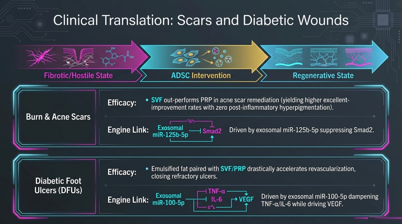
Smad2 is a signalling protein in the TGF-beta pathway, and that pathway is one of the best-characterised drivers of scar formation and fibrosis. A miRNA packaged in an exosome is a plausible delivery mechanism for interfering with it, because an exosome can carry a regulatory RNA into a recipient cell without the cell becoming a permanent graft. The slide presents that as the engine link, and the wording should be read as the proposed explanation for the observation rather than as an independently proven causal chain.
The diabetic foot ulcer panel follows the same two-part structure. It reports that emulsified fat paired with SVF or PRP drastically accelerates revascularisation, closing refractory ulcers, and it attaches a dual mechanism: exosomal miR-100-5p dampening TNF-alpha and IL-6 while driving VEGF. Here the intervention combines a structural graft with a cell fraction, and the reported endpoint is ulcer closure, which is a binary clinical outcome rather than an aesthetic grade.
| Panel | Reported effect on the slide | Proposed mechanism on the slide | Endpoint type |
|---|---|---|---|
| Burn and acne scars | SVF out-performs PRP in acne scar remediation, with higher excellent-improvement rates and zero post-inflammatory hyperpigmentation | Exosomal miR-125b-5p suppressing Smad2 | Graded improvement category |
| Diabetic foot ulcers | Emulsified fat paired with SVF or PRP drastically accelerates revascularisation, closing refractory ulcers | Exosomal miR-100-5p dampening TNF-alpha / IL-6 while driving VEGF | Ulcer closure |
The Signal Chain Each Panel Names
The two mechanism diagrams are the reason this slide is in a deck about fat graft biology rather than in a scar clinic's brochure. Both name an exosomal microRNA as the carrier of the signal, and both describe that signal as a change to the inflammatory environment rather than to the fat graft itself.
| Carrier named on the slide | Target named on the slide | Direction | Indication the slide pairs it with |
|---|---|---|---|
| Exosomal miR-125b-5p | Smad2 (in the TGF-beta / scar-formation pathway) | Suppression of the target | Burn and acne scars |
| Exosomal miR-100-5p | TNF-alpha and IL-6 (dampened) | Reduction of inflammatory cytokines | Diabetic foot ulcers |
| Exosomal miR-100-5p | VEGF (driven) | Increase in angiogenic signal | Diabetic foot ulcers |
The Three-State Frame, Read as a Table
The arrow across the top of the slide is the deck's own summary, and it is worth converting into rows because it shows the direction of travel the source material is arguing for.
| State | What the slide depicts | What the intervention is positioned to change |
|---|---|---|
| Fibrotic / hostile | Scar tissue, disrupted architecture, a signalling environment the slide illustrates with a molecular motif | Nothing at this stage — this is the starting condition |
| ADSC intervention | Cells and their secreted exosomes introduced into that environment | The inflammatory and signalling environment |
| Regenerative state | Reorganised tissue architecture and restored vascular supply in the illustration | The endpoint the slide labels as regenerative |
Two cautions belong with the slide rather than after it. First, neither indication is a cosmetic body-contouring procedure, and the reported endpoints — an improvement grade in scarring and closure of a refractory wound — are specific to those clinical settings. Second, "drastically" is the slide's adverb, not a measured effect size; the source material gives no percentage on this slide, and none should be inferred. What the slide does establish is the direction the field is moving in: from harvesting cells for volume to using their secreted signals for tissue repair.
Why the Deck Reaches Outside Cosmetics
Two properties make scarring and chronic wounds useful examples in a deck about fat grafting. The first is that both are conditions where the tissue environment itself is the problem. Scar tissue is dense, poorly vascularised and chronically inflamed, and a diabetic foot ulcer sits in tissue with compromised blood supply. Those are the conditions the deck's survival argument is about, stated at their extreme. If a graft and its cell fraction behave differently according to the recipient bed, then a hostile bed is precisely where the difference should be most visible.
The second is measurability. Ulcer closure is a binary endpoint that a clinician either observes or does not, and scar improvement can be graded against photographic standards, so both settings can produce outcomes recorded in categories rather than as impressions. That is what lets the slide attach an improvement grade to one panel and a closure endpoint to the other. The limitation is that neither endpoint transfers to elective body contouring, where the questions are volume, contour and skin quality rather than wound repair — and nothing on the slide suggests that it does.
Frequently Asked Questions
Is the slide saying SVF treats scars better than PRP?
The slide states that SVF out-performs PRP in acne scar remediation, yielding higher excellent-improvement rates with zero post-inflammatory hyperpigmentation. That is a comparison between two autologous preparations as reported in the source material, and the specific rate figures sit behind the study the slide is drawing on rather than on the slide itself.
What is the mechanism the slide gives for each indication?
For burn and acne scars the slide names exosomal miR-125b-5p suppressing Smad2, a protein in the TGF-beta signalling pathway. For diabetic foot ulcers it names exosomal miR-100-5p dampening TNF-alpha and IL-6 while driving VEGF.
Does this slide describe a cosmetic fat-grafting procedure?
No. Both panels sit outside cosmetic body contouring: acne and burn scarring, and refractory diabetic foot ulcers. The slide is using those settings to show what the cell fraction and its exosomes are proposed to do at the tissue level, not to describe an aesthetic indication.
This is published information, not medical advice — a board-certified surgeon must assess whether a procedure suits you.
Related Reading
- Why Transplanted Fat Survives — or Does Not
- Liposuction Risks and Complication Rates as Published
- Compression Garments and Lymphatic Massage After Liposuction
Estimating and comparing your own situation: risk and volume limits recovery timeline.