Four decisions the cannula slide actually encodes
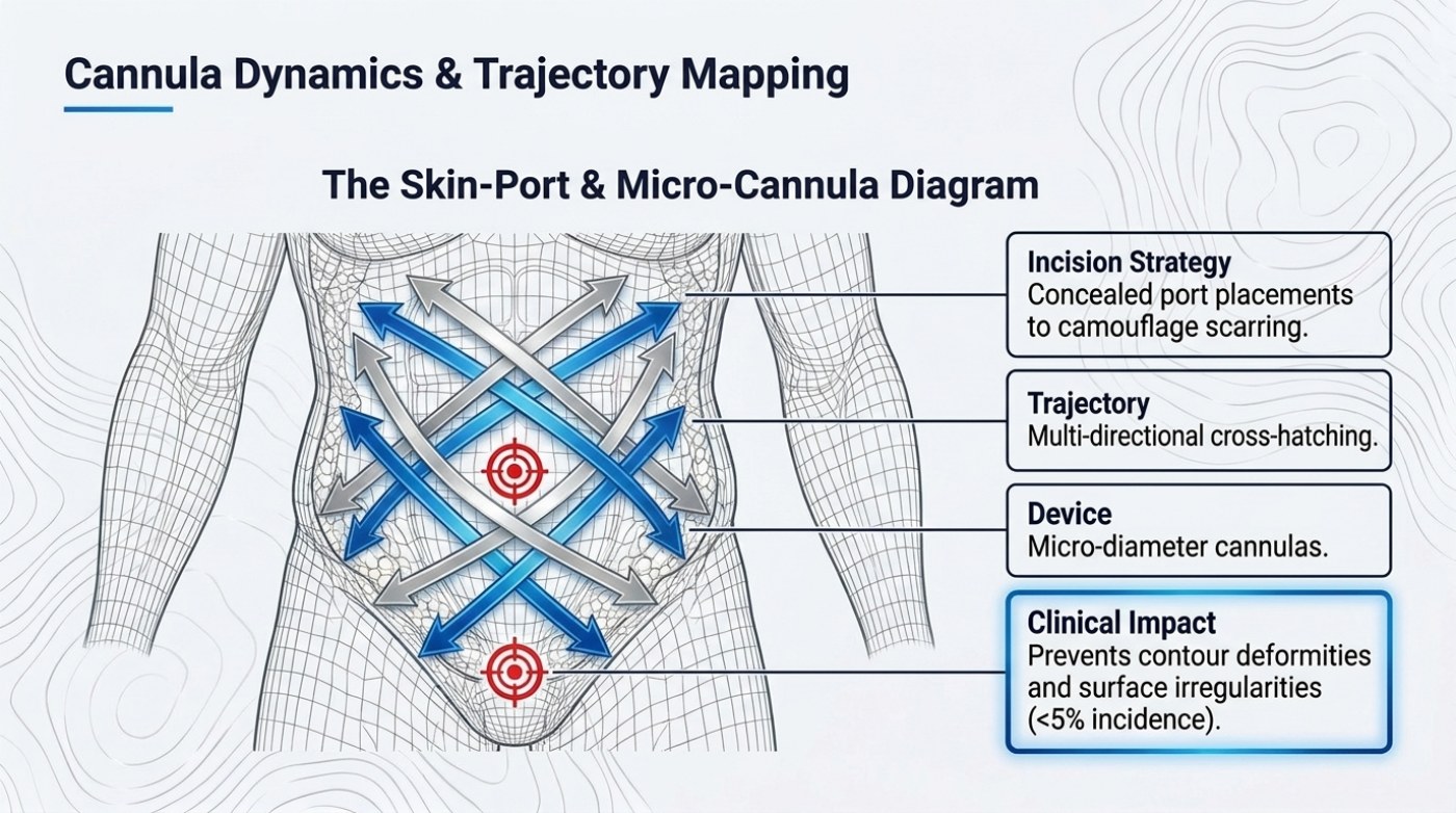
A cannula is not a vacuum cleaner. It is a direction. The instrument removes fat along the line it travels and leaves everything off that line untouched, which is why the same volume of aspirate can produce a smooth flank on one patient and a corrugated one on another. The deck slide “Cannula Dynamics & Trajectory Mapping” treats that geometry as four separate decisions rather than one technique: where the incision is made, which directions the instrument travels, how wide the instrument is, and what those three choices are supposed to protect. The slide presents them under a wireframe torso as a labelled diagram titled “The Skin-Port & Micro-Cannula Diagram”, with blue and grey trajectory lines crossing over the abdomen and a target marker drawn over the central contour. Most homeowners who read this are comparing facial liposuction options around Miami, FL, so what follows sticks to the details that change in practice.
The short version
- The cannula slide treats fat removal as four decisions: port placement, trajectory, instrument diameter and what they protect.
- Concealed port placement hides scarring while still giving the cannula a straight working line into the target area.
- Multi-directional cross-hatching runs passes at differing angles so residual fat is not left in parallel ridges.
- Micro-diameter cannulas remove smaller increments, giving finer control near the skin where the fat layer is thinnest.
- Published pooled contour deformity rates run 2.35% in one 39-study analysis and 2% in a second review.
Port placement is both a scarring decision and a reach decision
The first callout box reads “Incision Strategy — concealed port placements to camouflage scarring.” A port is the small incision the cannula enters through, and where it sits determines two things at once. Cosmetically, the scar should end up somewhere a waistband, a fold or a natural crease already hides. Mechanically, the port has to give the cannula a straight working line into the tissue being treated; from a badly placed port the instrument reaches the target at an angle that limits the directions available to it. The two constraints pull against each other, and the compromise is made before a single pass is taken. Our published guide to abdominal etching describes the same planning step from the other end, with ports placed deliberately for the anterior abdomen, the flanks and the pectorals so that a fine cannula can reach each groove without crossing fat that is being left in place.
Cross-hatching is what turns one direction into a plane
The second box, “Trajectory — multi-directional cross-hatching,” describes the answer to a straightforward problem. If every pass runs along the same axis, the residual fat forms ridges aligned with that axis, and the skin heals down onto them. Cross-hatching breaks the pattern by running successive passes at different angles within the same treatment zone, so the remaining fat is distributed between intersecting tracks instead of left in parallel ribbons. The slide's diagram shows exactly this: silver lines and blue lines crossing the abdomen at opposing angles rather than fanning out from a single direction. The technique costs time — more passes, more instrument handling — and it is the part of the plan that a patient can ask about directly, because it is decided rather than improvised.
Why micro-diameter instruments change the finish
The third element is the device itself: “micro-diameter cannulas.” Smaller bore instruments remove smaller increments of fat with each pass, which gives the operator finer control near the surface where the fat layer is thin and the margin for error is measured in millimetres. Published work on cannula and needle diameter and adipocyte survival has repeatedly linked instrument size to how cells are handled, which is why a definition-focused case typically uses one instrument set for bulk work in the deep layer and a finer set for the superficial pass. Our etching guide sets the same two-pass structure out in layer terms: the deep compartment is emulsified and aspirated in bulk with larger cannulas to establish circumference, and the superficial layer is selectively thinned with fine cannulas to trace grooves. The slide's four elements only make sense together, because a fine cannula in the wrong trajectory produces the same ridge in a smaller size.
What the “<5% incidence” figure is and is not
The fourth box states the clinical impact the other three are bought for: this combination “prevents contour deformities and surface irregularities (<5% incidence).” The figure belongs to the source material for this deck, not to a named study, so it is worth setting beside what is actually published on the same endpoint. Pooled meta-analysis has reported contour deformity or irregularity at 2.35% in one 39-study analysis and 2% in a second review of isolated aesthetic liposuction. A systematic review of contemporary techniques reported a far wider spread by modality — ultrasound-assisted liposuction pooled at roughly 0.12% for contour irregularities against roughly 3.36% for traditional suction-assisted liposuction — and older studies relying on surgeon self-reporting have cited figures as high as 9%. That spread, not any single number, is the useful finding: irregularity is the most commonly reported complication in pooled datasets and the one technique choices move most.
The four decisions, side by side
| Element on the slide | What it means in the operative plan | What it is intended to protect |
|---|---|---|
| Incision strategy — concealed port placements | Ports sited where a scar is hidden, while still giving the cannula a workable line into the target zone | Scar visibility and the range of trajectories available inside the treatment area |
| Trajectory — multi-directional cross-hatching | Successive passes at differing angles through the same zone instead of parallel single-plane passes | Ridging and step-off where the remaining fat heals into a visible pattern |
| Device — micro-diameter cannulas | Smaller-bore instruments that remove finer increments, usually with a separate set for the superficial pass | Control near the skin surface, where the fat layer is thinnest |
| Clinical impact — <5% incidence | The source material’s stated target for contour deformity and surface irregularity | The visible finish of the contour rather than the volume removed |
Reported irregularity rates: the slide beside the published record
| Figure | Value | Where it comes from |
|---|---|---|
| Deck benchmark for contour deformities and surface irregularities | <5% incidence | Slide source material for this deck; not attributed to a named study |
| Contour deformity / irregularity, pooled | 2.35% | Comerci et al., 39 studies, 29,368 patients |
| Contour deformity / irregularity, pooled | 2% | Aljerian et al., review of isolated aesthetic liposuction |
| Comparative modality pooling | About 0.12% ultrasound-assisted vs about 3.36% suction-assisted | Systematic review of contemporary techniques |
| Older self-reported series | Up to 9% | Studies relying on surgeon self-reporting |
What a patient can check in the consultation
Nothing on this page is a clinical instruction, but the slide does translate into questions with observable answers. Which ports are planned, and where each of them sits on the body. Whether the plan involves cross-hatching at all, or a single-plane approach. Whether two instrument sizes are used and which layer each is for. And what the practice’s own reported rate of contour irregularity and revision is, against the published pooled figures above. Those four answers describe the operative geometry more precisely than any description of how much fat will be removed, because the geometry is what the patient looks at afterwards.
Where this sits inside the wider protocol
The deck places cannula dynamics between tumescent infiltration — which shrinks vessels, separates fat from connective tissue and makes the superficial pass feasible — and the energy adjuncts that address what remains: skin retraction after the fat is gone. Each stage assumes the one before it. A cross-hatched trajectory in an untreated field bleeds more, and skin tightening over an uneven surface produces a tight uneven surface. Volume limits and thromboembolism prophylaxis sit alongside, for reasons that have nothing to do with contour and everything to do with how long the case runs and how much fluid shifts.
FAQ: cannula dynamics and trajectory
Does cross-hatching make recovery harder?
It changes the pattern of bruising rather than the substance of recovery. Published accounts of liposuction recovery describe peak swelling and bruising in the first days, then progressive resolution over weeks, with compression garments worn continuously in the early period. More passes through the same zone involve more instrument handling, and the published record ties bruising and ecchymosis to that handling; post-operative lymphatic taping has been reported to speed resolution of abdominal ecchymosis after body contouring. What the deck describes is a trade: extra operative time and instrument handling in exchange for a surface less likely to heal into visible ridges.
Why would two different cannula sizes be used in one operation?
Because the two layers have different tolerances. Bulk removal in the deep compartment tolerates a larger instrument and benefits from its speed; the superficial layer directly under the skin does not, and published work on instrument diameter and adipocyte survival links bore size to how cells are handled. The site’s published etching guide describes the same split: larger cannulas for the deep pass that sets the overall silhouette, fine cannulas for the superficial pass that traces muscle grooves.
Is a “<5% incidence” figure a guarantee?
No. The figure on the slide is the source material’s stated benchmark for contour deformity and surface irregularity, and it appears on a slide whose overall argument is that benchmarks only hold when the whole protocol is followed. Published pooled rates for the same endpoint range from below 1% to above 9% depending on technique and how the endpoint is defined, and a complication rate is a population statistic, not a prediction for one person. Rates are reported about groups; suitability is a separate question that requires an examination.
This is published information, not medical advice — a board-certified surgeon must assess whether a procedure suits you.
Related reading
Three published guides on this site cover adjacent parts of the same protocol:
- High-Definition Liposuction: How Muscle Etching Actually Works — the two-layer sequence this instrument set serves
- Liposuction Risks and Complication Rates as Published — the pooled rates behind contour-irregularity claims
- How Much Fat Can Be Removed in One Liposuction Session? — how volume decisions interact with technique