Between the moment a fat graft is placed and the moment new capillaries reach it, the transferred cells are living without a blood supply. The presentation calls this the avascular window and puts its length at one to three days. During that period the graft's cells are working through whatever energy they have stored, in tissue that is short of oxygen and short of nutrients, while the surrounding environment is at its most inflammatory. The third mechanism on the deck, Mechanism 3: Anti-Apoptosis, describes what the enriched cells are said to do about that.
The short version
- Anti-apoptosis is the mechanism described for keeping transferred fat cells alive through a one to three day avascular window.
- The slide's cascade pairs upregulation of Bcl-2, an anti-apoptotic protein, with direct suppression of the caspase pathways.
- Apoptosis is programmed cell death, an orderly self-dismantling sequence distinct from the uncontrolled breakdown that follows overwhelming injury.
- The slide lists lipid liquefaction as the second post-operative outcome the mechanism is intended to avoid.
- A three-patient comparison found adipocyte counts 47% higher immediately after harvest at minus 250 mmHg than at minus 760 mmHg.
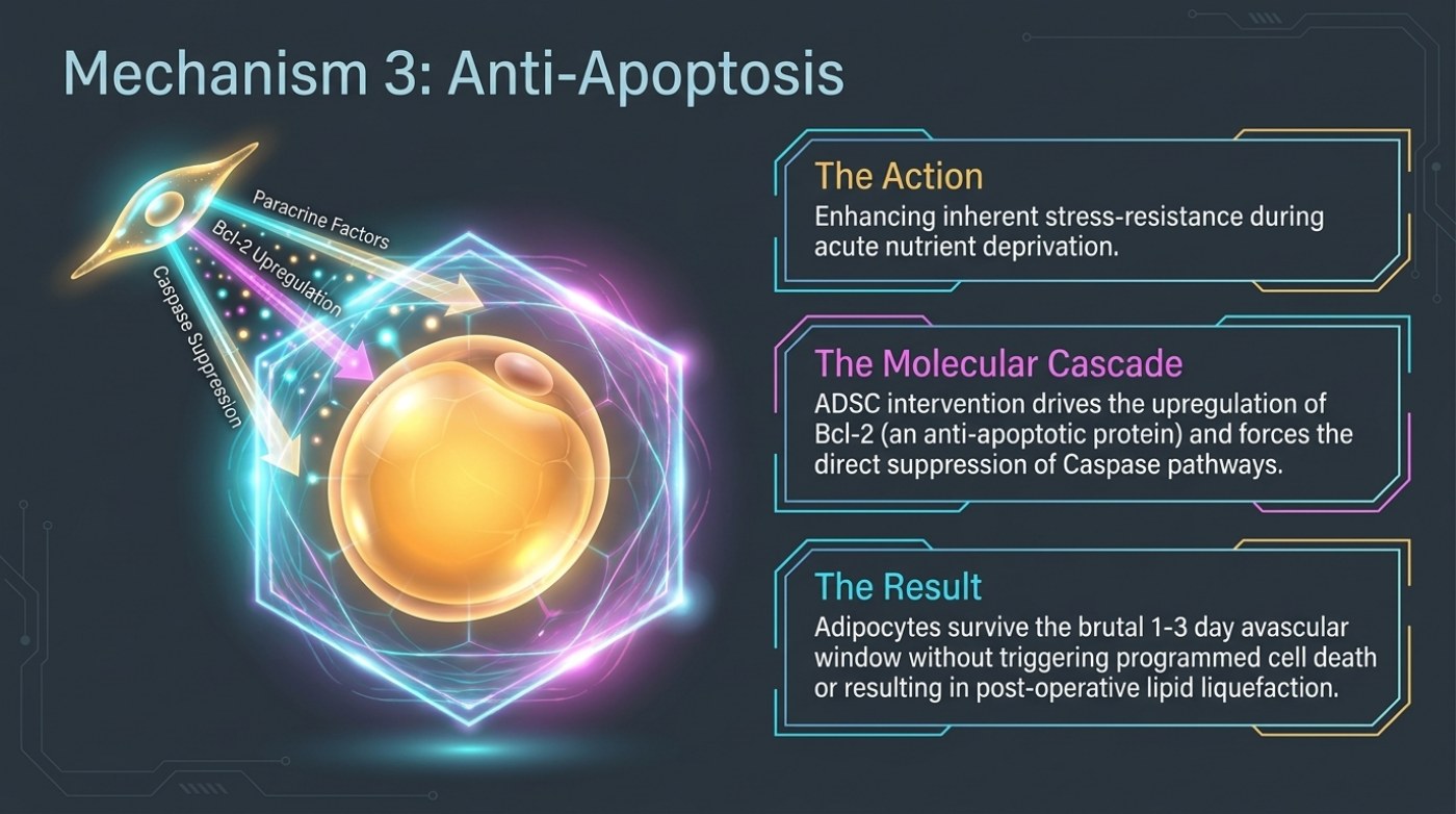
The slide's left panel shows a golden fat cell enclosed in a translucent shield — a visual shorthand for protection — with an ADSC above it sending three labelled arrows inward. The arrows read "Paracrine Factors", "Bcl-2 Upregulation" and "Caspase Suppression". To the right, three bordered panels carry the explanation: The Action, The Molecular Cascade and The Result.
The three panels, and what sits between them
The Action panel is about tolerance rather than growth: enhancing inherent stress-resistance during acute nutrient deprivation. The Molecular Cascade panel names the machinery — ADSC intervention drives the upregulation of Bcl-2, described on the slide as an anti-apoptotic protein, and forces the direct suppression of caspase pathways. The Result panel states the intended consequence: adipocytes survive the brutal one-to-three-day avascular window without triggering programmed cell death or resulting in post-operative lipid liquefaction.
What connects the panels is a fairly simple idea. A cell that is deprived of oxygen and nutrients does not simply run out of fuel and stop; it can be actively triggered to dismantle itself through a controlled sequence that the cell runs on purpose. That sequence is apoptosis, and the slide's argument is that the enriched cells push the machinery of that sequence in the other direction for as long as it takes the graft to be reconnected to a blood supply.
| Panel | Slide wording | What it describes | What it leaves open |
|---|---|---|---|
| The Action | Enhancing inherent stress-resistance during acute nutrient deprivation | Increasing how much deprivation a transferred fat cell can tolerate | How much resistance, for how many cells, in which recipient sites |
| The Molecular Cascade | ADSC intervention drives the upregulation of Bcl-2 and forces the direct suppression of caspase pathways | The intracellular machinery named as responsible | Any dose, delivery method or timing detail |
| The Result | Adipocytes survive the brutal 1-3 day avascular window without triggering programmed cell death or resulting in post-operative lipid liquefaction | The window the mechanism is aimed at, and two of the failure modes it is meant to avoid | Any measured retention figure for a given preparation |
The vocabulary, term by term
Apoptosis is programmed cell death — an orderly, energy-dependent process in which a cell disassembles itself rather than rupturing. It is different from necrosis, which is the uncontrolled breakdown that follows overwhelming injury. The slide's wording is specific: it describes adipocytes surviving "without triggering programmed cell death", which places the mechanism on the apoptosis side of that distinction, and separately names lipid liquefaction as the second outcome avoided.
Bcl-2 is a protein that sits on the anti-apoptotic side of the balance; the slide calls it an anti-apoptotic protein and describes its upregulation as one half of the cascade. The caspases are a family of enzymes that carry out the dismantling steps of apoptosis, and the slide describes their pathways as being directly suppressed. Paracrine factors — the third label on the graphic — are signals released by a cell that act on neighbouring cells rather than on itself, which is the same class of communication described on the angiogenesis and immunomodulation slides.
| Term on the slide | How the slide uses it | Plain-language note |
|---|---|---|
| Apoptosis | Programmed cell death, named as the outcome to be avoided | A controlled self-dismantling sequence the cell runs, distinct from necrosis |
| Avascular window (1-3 days) | The period the slide says adipocytes must survive | The gap before the graft is reconnected to a blood supply — the same gap described as the ischemic problem elsewhere in the deck |
| Bcl-2 | Upregulated; described as an anti-apoptotic protein | One side of the intracellular balance that decides whether a stressed cell survives |
| Caspase pathways | Directly suppressed | The enzyme cascade that executes the dismantling steps of apoptosis |
| Paracrine factors | One of the three arrows aimed at the fat cell | Signals released by one cell that act on its neighbours |
| Lipid liquefaction | Named as the second post-operative outcome avoided | The breakdown of dead fat cells into liquid lipid — the process behind oil-cyst formation and irregularity |
Why the two halves of the cascade matter together
The slide presents the cascade as two coordinated moves: raise the protection, lower the execution. That pairing is what makes the mechanism legible. Raising Bcl-2 without suppressing the caspase side would leave a cell that is more resistant but still wired to dismantle itself; suppressing caspases without the protective side would remove the execution step without addressing why the cell was being pushed toward it. The slide's claim is that the enriched cells do both, which is why it describes the result as cells that survive the window rather than cells that merely delay their failure.
It is also worth noting what the slide does not say. It gives no percentage of cells saved, no measurement of retention, and no indication that the effect is uniform across a graft. Our guide to why transplanted fat survives puts the underlying stakes plainly: published reviews describe volume loss across conventional grafting of between 30% and 80%, and the material behind this site describes as much as 80% of a conventional transfer being reabsorbed. Anti-apoptosis is offered as one of the reasons that number is as wide as it is, not as a number of its own.
The harvest still sets the ceiling
There is a practical consequence to the slide's emphasis on what happens inside the cell. If survival depends on the state of the adipocytes that arrive, then the way they were removed matters before enrichment is even considered. The site's fat-graft survival guide reports a three-patient comparison in which adipocyte counts immediately after harvest were 47% higher at −250 mmHg than at −760 mmHg, with cell viability still significantly higher at day seven. It also records that the source material for this site describes harvesting at roughly 400 mmHg with cannulas near 3 mm, while cautioning that the published comparison used a different pair of pressures, so the exact figure belongs in a question rather than an assumption. Cannula diameter is described in the same guide as a documented determinant of fat-cell survival, because shear forces rise as the instrument narrows.
| Variable | Published figure or description | Where it comes from |
|---|---|---|
| Vacuum pressure and adipocyte count | 47% higher count immediately after harvest at −250 mmHg versus −760 mmHg, with better day-seven viability | Three-patient comparison reported in our fat-graft survival guide |
| Harvesting setting described in the source material | Roughly 400 mmHg with cannulas near 3 mm | Source material for this site, as quoted in the same guide |
| Volume loss in conventional grafting | Reported range 30% to 80%; up to 80% described in the source material | Published fat-grafting reviews, as summarised in the same guide |
| Cell purity by preparation route | Uncultured stromal vascular fraction roughly 1-10% stem or stromal cells; culture-expanded cells greater than 90% purity | Same guide, describing the two cell-preparation routes |
| Overall complication rate, pooled | 2.62% in one meta-analysis; 12% in another; 1.16% in a 69,424-patient registry | Our guide to published complication rates |
The recovery question this mechanism raises
If a graft's first days are the deciding days, then what happens to the treated area in those days is not passive. Our guide to recovery after liposuction sets out the published sequence — swelling peaks, bruising fades, the area firms up and then softens — and notes that the timeline is predictable in shape and variable in pace. Compression, positioning and activity instructions exist in that frame. The slide describes a cellular mechanism, not a recovery protocol, and no part of it should be read as a substitute for the instructions a treating surgeon gives about the days immediately after a procedure.
Frequently asked questions
Is anti-apoptosis the same as keeping the fat alive?
It is one of the routes described for keeping more of it alive. The slide frames it as protecting the transferred adipocytes through the avascular window so they are still present when blood supply returns. The other mechanisms in the same deck — new vessel growth, the immune-environment shift and mitochondrial transfer — address the same window from different angles.
Does surviving the window mean the fat stays?
No. Clearing the avascular window is a precondition for retention, not a guarantee of it. Grafts are vulnerable throughout the remodelling period that follows, and our fat-graft survival guide notes that published reviews describe wide variability between patients and between recipient sites, which is why it recommends treating any figure as a range and a plan rather than a promise.
What does "lipid liquefaction" actually refer to?
It is the slide's term for what happens when fat cells die and their lipid content breaks down into liquid rather than being cleared cleanly. The practical consequences discussed in the literature are collections and irregularities within the grafted area. The slide names it as the outcome the anti-apoptotic mechanism is intended to avoid.
Related reading
Why Transplanted Fat Survives — or Does Not — pressure, cannula diameter, purification, layering and enrichment in one place.
How Much Fat Can Be Removed in One Liposuction Session? — the published volume thresholds, which bear directly on how thickly grafted tissue sits.
Liposuction Recovery Week by Week — the published sequence of swelling, firmness and softening after a procedure.
This is published information, not medical advice — a board-certified surgeon must assess whether a procedure suits you.