The days immediately after fat is transferred into a recipient site are not quiet. Tissue has been disrupted, small vessels have been torn, and the body's first response is inflammatory — a wave of signalling that recruits immune cells to the injury. That response is normal and necessary, but in the setting of a fat graft it works against the transferred tissue as well as for it. The second mechanism in the presentation, titled Mechanism 2: Immunomodulation, describes the way enriched adipose-derived cells are described as steering that response rather than simply enduring it. Most homeowners who read this are comparing body contour services options around Miami, FL, so what follows sticks to the details that change in practice.
The short version
- Immunomodulation is the second mechanism, describing how enriched cells are said to steer the body's inflammatory response to a fat graft.
- The slide shows a shift from the pro-inflammatory M1 macrophage state to the anti-inflammatory, tissue-healing M2 state.
- Adipose-derived stem cells are shown releasing IL-10, TGF-beta and PGE2 as the signal driving that transition.
- One panel describes an anti-fibrotic route in which exosomal miR-125b-5p suppresses Smad2 in fibroblasts, halting collagen over-deposition.
- The slide frames the action as quieting the post-surgical inflammatory storm during the critical acute phase.
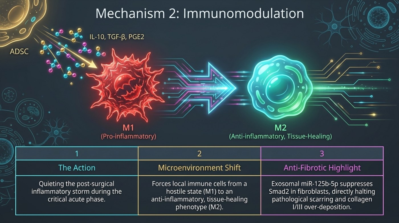
The slide's central graphic is a transition, not a process chain. On the left is a red, spiked cell labelled M1 (Pro-inflammatory). On the right is a smooth, green-teal cell labelled M2 (Anti-inflammatory, Tissue-Healing). A large arrow connects them. Above the M1 cell, an ADSC is shown releasing a cluster of coloured molecules labelled IL-10, TGF-β and PGE2 — the signal that the slide says drives the transition.
M1 and M2: two states of the same cell
Macrophages are immune cells that change what they do depending on the signals around them. The M1 and M2 labels are a shorthand for two ends of that spectrum. The slide calls M1 "pro-inflammatory" and M2 "anti-inflammatory, tissue-healing". The distinction matters to a graft because the two states are associated with different downstream behaviour: an environment dominated by inflammatory signalling is one in which the transferred tissue contends with more stress, while an environment shifted toward repair is the one in which new tissue can be organised rather than broken down.
It is worth being precise about what the slide does and does not claim. It does not say that inflammation is suppressed altogether, and it does not describe a drug, a dose or an external treatment. It describes a local shift in the balance of immune-cell states, prompted by factors released by the enriched cells mixed into the graft.
| Feature | M1 (as labelled on the slide) | M2 (as labelled on the slide) |
|---|---|---|
| Slide label | Pro-inflammatory | Anti-inflammatory, tissue-healing |
| Position in the diagram | Starting state, left of the transition arrow | Resulting state, right of the transition arrow |
| What drives the change on the slide | ADSC release of IL-10, TGF-β and PGE2 | Same signal, described as forcing the shift away from the M1 state |
| Described effect on the graft environment | Hostile post-surgical inflammatory state during the acute phase | Microenvironment shifted toward repair, with less collagen over-deposition described |
| Where it appears again | — | The breast-reconstruction slide cites M2 macrophage polarisation as part of how fat grafting is described as acting on radiation-induced fibrosis |
The three panels the slide uses to explain the action
Below the graphic, the slide carries a three-column panel. The first column, "The Action", describes quieting the post-surgical inflammatory storm during the critical acute phase. The second, "Microenvironment Shift", states that the intervention forces local immune cells from a hostile state (M1) to an anti-inflammatory, tissue-healing phenotype (M2). The third, "Anti-Fibrotic Highlight", is the most specific of the three: exosomal miR-125b-5p suppresses Smad2 in fibroblasts, which the slide describes as directly halting pathological scarring and collagen I/III over-deposition.
Read together, the three panels describe a single trajectory with a long tail. The acute phase is the emergency — the first days, when the graft is avascular and the local environment is at its most hostile. The microenvironment shift is the change in state that outlasts the acute phase. The anti-fibrotic panel is the part that looks furthest ahead, into how the tissue remodels over the weeks and months that follow, and specifically into the scarring response that can accompany it.
| Panel on the slide | Slide wording | What it implies for the graft | What it does not state |
|---|---|---|---|
| 1 — The Action | Quieting the post-surgical inflammatory storm during the critical acute phase | The most stressful window is the one the mechanism is aimed at | Any timing, dose or administration detail |
| 2 — Microenvironment Shift | Forces local immune cells from a hostile state (M1) to an anti-inflammatory, tissue-healing phenotype (M2) | A change in the recipient bed itself, not only in the transferred cells | Any quantification of the shift, or of how long it persists |
| 3 — Anti-Fibrotic Highlight | Exosomal miR-125b-5p suppresses Smad2 in fibroblasts, halting pathological scarring and collagen I/III over-deposition | A described route by which the remodelling phase is influenced | Any measured scar or fibrosis outcome in humans from this slide alone |
The molecular names, explained without the jargon
IL-10 (interleukin-10), TGF-β (transforming growth factor beta) and PGE2 (prostaglandin E2) are the three factors the slide shows being released by the ADSC. Each has a long history in immunology as a modulator of inflammation, and the slide uses all three as a group label for the signal that precedes the M1-to-M2 transition.
The anti-fibrotic panel introduces two further terms. Smad2 is a protein inside fibroblasts that relays TGF-β-family signals into changes in gene activity; suppressing it is the step the slide identifies as interrupting the scarring pathway. Collagen I/III refers to the two collagen types most involved in scar tissue, and "over-deposition" is the slide's phrase for the excess that produces a stiff, thickened result. The mechanism is delivered, in the slide's telling, by an exosome — a small vesicle — carrying miR-125b-5p rather than by a live cell acting directly on the fibroblast.
| Term | Role as described on the slide | Plain-language note |
|---|---|---|
| IL-10, TGF-β, PGE2 | Released by the ADSC; the signal shown preceding the M1-to-M2 transition | Three named mediators of the inflammatory response, grouped as one signal on the slide |
| M1 macrophage | Pro-inflammatory state; the starting point of the transition | The state associated with the immediate post-injury reaction |
| M2 macrophage | Anti-inflammatory, tissue-healing state; the endpoint of the transition | The state associated with repair and remodelling, cited again on the breast-reconstruction slide |
| Exosomal miR-125b-5p | Carried by an ADSC-derived vesicle; suppresses Smad2 in fibroblasts | A microRNA payload, not a living cell — the same class of delivery described on the angiogenesis slide |
| Smad2 | Suppressed by miR-125b-5p; the relay the slide says is interrupted | An intracellular signalling protein in fibroblasts, in the TGF-β family of pathways |
| Collagen I/III over-deposition | Described as halted by the same mechanism | The excess collagen that produces stiff, thickened scar tissue |
Where inflammation meets the published outcome data
Our guide to liposuction risk and volume notes a pattern in the published complication data that is consistent with the slide's emphasis on the acute phase: the complications that dominate are the ones involving fluid and tissue response rather than catastrophic events. Seroma — a collection of lymphatic fluid in the space where fat was removed — is the most common complication in the ASPS TOPS analysis of more than 4,500 patients, and above the five-litre aspirate line it is responsible for almost the entire rise in complication rate from 1.1% to 3.7%. Contour deformity, at 2.35% in one pooled meta-analysis and 2% in another, is the most common finding in every pooled dataset. Those figures describe liposuction procedures generally, not fat grafting specifically, and they are not evidence that immune modulation changes any of them. They are the context in which a mechanism like this is worth understanding.
The site's fat-graft survival guide records the same mechanism from the clinical side, listing a shift of macrophages toward the anti-inflammatory M2 phenotype among the biological events described in the source material, alongside pro-angiogenic growth-factor secretion and mitochondrial transfer. That guide also states plainly why no mechanism should be converted into a promise: published systematic reviews report retention improvements from cell enrichment while noting that study designs, cell preparation and dose vary enough between them that a single percentage does not transfer to an individual patient.
Frequently asked questions
Is immunomodulation the same as being immunosuppressed?
No. The slide describes a local change in the balance of immune-cell states inside the graft and its immediate surroundings, not a systemic suppression of immune function. Nothing in the material describes a drug or a treatment given to alter immunity as a whole.
Does this mean scarring and fibrosis are prevented?
The slide describes exosomal miR-125b-5p suppressing Smad2 in fibroblasts and halting collagen I/III over-deposition as a mechanism. That is a description of a laboratory-level pathway, not a measured clinical scar result, and it should not be read as a claim about how any particular person's tissue will heal.
Why does the same mechanism appear on more than one slide?
Because it is cited twice in the presentation for different purposes. On this slide it explains the acute inflammatory phase; on the breast-reconstruction slide it is cited as the route by which fat grafting is described as acting on radiation-induced fibrosis. The four mechanisms described in this deck overlap by design rather than sitting in separate compartments.
Related reading
Why Transplanted Fat Survives — or Does Not — the harvest and enrichment variables that set up the local response.
Compression Garments and Lymphatic Massage After Liposuction — what the published guidance says about managing swelling and fluid in the recovery period.
Liposuction Risks and Complication Rates as Published — seroma, contour deformity and the rest, with a source behind every number.
This is published information, not medical advice — a board-certified surgeon must assess whether a procedure suits you.