A fat graft has no blood supply at the moment it is placed. Every cell in the transferred tissue lives on diffusion from the surrounding bed until new capillaries reach it, and the volume that survives is decided largely by how quickly that happens. Our guide to why transplanted fat survives sets out the arithmetic: published reviews put the volume lost in conventional grafting somewhere between 30% and 80%, and the source material behind this site describes as much as 80% of a conventional transfer being reabsorbed. The first of the four biological mechanisms the presentation sets out is aimed squarely at that gap. It is titled Mechanism 1: Rapid Angiogenesis, and its subject is the growth of a new local blood supply.
The short version
- Rapid angiogenesis is the first of four mechanisms described for keeping transplanted fat alive before new vessels arrive.
- Adipose-derived stem cells release VEGF-A, FGF2 and Ang-1 directly, triggering endothelial cell proliferation and tube formation in the recipient bed.
- A second route uses ADSC-derived exosomes carrying miR-21-5p, which target the NOTCH1, DLL4 and VEGFA axis.
- The slide describes rapid neovascularisation as preventing the central graft necrosis typical of large-volume fat transfers.
- An ASPS TOPS analysis of more than 4,500 patients found complications rising from 1.1% to 3.7% above five litres.
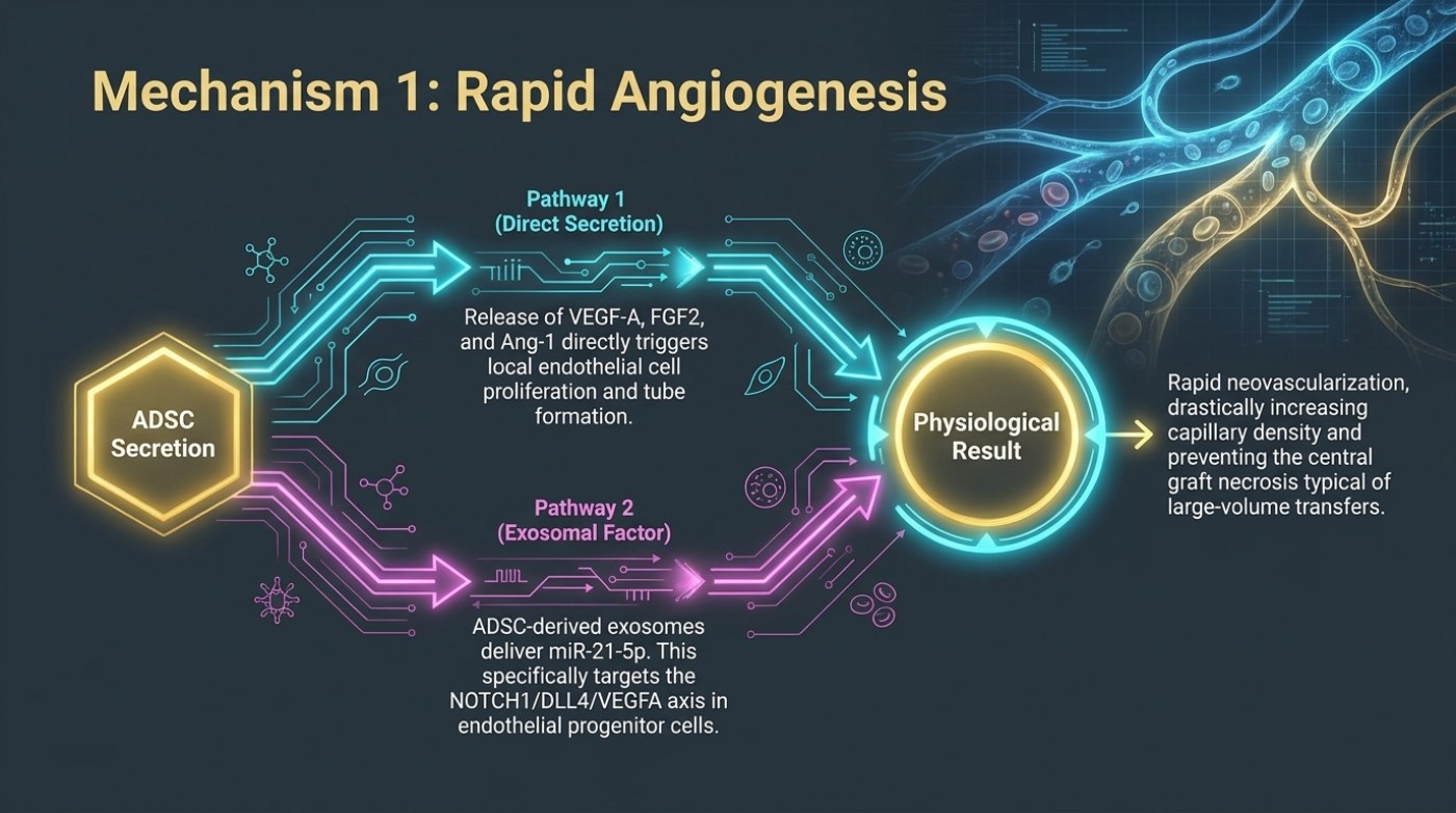
The slide is drawn as a two-branch circuit. On the left sits the adipose-derived stem cell, labelled "ADSC Secretion". Two coloured pathways run from it to the right, where a single circular node is marked "Physiological Result". Above the node the slide states the consequence: rapid neovascularisation, drastically increasing capillary density and preventing the central graft necrosis typical of large-volume transfers. The two branches differ only in how the signal travels.
The two pathways the slide separates
Pathway 1 is labelled "Direct Secretion". In the slide's wording, the release of VEGF-A, FGF2 and Ang-1 directly triggers local endothelial cell proliferation and tube formation. Nothing is packaged and nothing is processed; the factors are released into the immediate surroundings of the graft and act on the endothelial cells that line existing capillaries in the recipient bed. The endpoint described is a sprouting response — new tubes forming where there were none, within reach of the transplanted tissue.
Pathway 2 is labelled "Exosomal Factor" and takes a different route. ADSC-derived exosomes deliver miR-21-5p, and the slide states that this specifically targets the NOTCH1/DLL4/VEGFA axis in endothelial progenitor cells. Here the signal is not a free-floating protein but a small vesicle carrying a microRNA, and its described target is not only the mature endothelial cells of the local bed but the progenitor cells that can be recruited into building new vessel lining. Two very different pieces of cell biology are being presented as one outcome.
| Route | What the slide says is released | Named target | Described endpoint |
|---|---|---|---|
| Pathway 1 — direct secretion | VEGF-A, FGF2 and Ang-1 released from the ADSC | Local endothelial cells | Endothelial cell proliferation and tube formation |
| Pathway 2 — exosomal factor | ADSC-derived exosomes carrying miR-21-5p | The NOTCH1/DLL4/VEGFA axis in endothelial progenitor cells | Vessel formation via a progenitor-cell route |
| Shared outcome (slide label) | — | Physiological result | Rapid neovascularisation, higher capillary density, prevention of central graft necrosis in large-volume transfers |
The growth factors named, in plain terms
VEGF-A is vascular endothelial growth factor A, the best-known signal for new vessel growth; FGF2 is fibroblast growth factor 2; Ang-1 is angiopoietin-1, which acts on the stabilisation and maturation side of vessel formation rather than on the initial sprout alone. The slide groups all three under a single action — triggering proliferation and tube formation in local endothelial cells — and gives no concentrations, no ratios and no delivery protocol. That omission is worth noticing rather than filling in. The presentation is describing a biological mechanism, not a recipe, and no part of it should be read as a specification for how a graft ought to be prepared.
What the mechanism does explain is why graft geometry matters as much as graft volume. If survival depends on capillaries arriving, then the distance any cell sits from the nearest perfused tissue is a design variable. Thin, layered placement keeps more of the graft within diffusion reach of the bed, which is the same logic our fat-graft survival guide describes when it distinguishes a single bolus from multi-layer placement.
| Named factor | Described role in the source material | Why the graft's outcome depends on it | Reasonable question to ask |
|---|---|---|---|
| VEGF-A | Released by ADSCs; triggers endothelial cell proliferation and tube formation | Starts the sprouting response that will perfuse the transferred fat | "How is the graft placed to keep it thin enough to be reached?" |
| FGF2 | Released alongside VEGF-A in the direct-secretion pathway | Contributes to the same proliferation-and-tube-formation endpoint | "Is any growth-factor or cell enrichment being added, and how is it prepared?" |
| Ang-1 | Named in the same release group as VEGF-A and FGF2 | Associated with vessel maturation rather than sprouting alone | "What is the plan if retention falls short — is a second stage contemplated?" |
| Exosomal miR-21-5p | Delivered by ADSC exosomes to the NOTCH1/DLL4/VEGFA axis in endothelial progenitor cells | Describes a second, vesicle-mediated route to the same endpoint | "Is the enrichment minimally manipulated tissue or a cultured cell product?" |
What the exosomal pathway adds to the picture
The exosomal branch is where the presentation's language becomes most specialised, and it is also where the wider literature on cell enrichment is most active. An exosome is a small vesicle released by a cell; a microRNA such as miR-21-5p is a short regulatory RNA carried inside it. The NOTCH1/DLL4/VEGFA axis named on the slide is a signalling relationship between a receptor (NOTCH1), its ligand (DLL4) and vascular endothelial growth factor A — the point being that a vesicle-borne signal can arrive at a vessel-forming cell and change what that cell does next.
Our guide to fat-graft survival describes the same theme from the clinical side: pro-angiogenic growth-factor secretion from enriched cells, a shift of macrophages toward the anti-inflammatory M2 phenotype, and mitochondrial transfer between cells through tunnelling nanotubes. It also notes how differently the two cell-preparation routes are regulated. Uncultured stromal vascular fraction is a fresh cell mix in which roughly 1–10% of cells are stem or stromal cells; culture-expanded adipose-derived stem cells are grown under GMP conditions to greater than 90% purity. The guide reports retention gains above 60% attributed to culture-expanded cells in the source material, while cautioning that published systematic reviews show retention improvements in which study designs, cell preparation and dose vary enough that a single percentage does not transfer to an individual patient. Angiogenesis is the mechanism most often invoked for that difference — and it is still a mechanism, not a measured guarantee.
Why the timing of revascularisation is the whole point
The slide's own justification for calling this mechanism important sits in its last line: rapid neovascularisation is described as preventing the central graft necrosis typical of large-volume transfers. The phrase "central" is the clue. A large collection of grafted fat has a core that is furthest from any perfused tissue, and that core is what suffers first when the vessel response is slow. The site's published guide to liposuction risk and volume notes a similar shape in the outcome data for high-volume procedures: in an ASPS TOPS analysis of more than 4,500 patients, the overall complication rate rose from 1.1% below the five-litre aspirate line to 3.7% above it, with the increase attributed almost entirely to seroma. Those figures describe liposuction generally rather than graft take, but the underlying theme — volume and vascularity are in tension — is the same.
| Published figure | Rate | Source, as cited on this site |
|---|---|---|
| Overall complication rate, pooled meta-analysis | 2.62% (39 studies, 29,368 patients) | Comerci et al., Aesthetic Surgery Journal, 2024 |
| Overall complication rate, second meta-analysis | 12% (60 studies, 21,776 patients) | Aljerian et al. |
| Overall complication rate, liposuction alone | 1.16% (69,424-patient registry) | Saad et al. |
| Complication rate below and above 5 L aspirate | 1.1% versus 3.7%, mostly seroma | ASPS TOPS analysis, more than 4,500 patients |
| Contour deformity — the most common finding in every pooled dataset | 2.35% (Comerci); 2% (Aljerian) | Both meta-analyses |
What "rapid angiogenesis" does not promise
A mechanism slide is not a result slide. Nothing in this material says a graft will take, or that a given technique produces a given retention. What it does say is that revascularisation is the variable that separates a graft that keeps its volume from one that does not, and that the same variable can be influenced before the fat ever leaves the body — by vacuum pressure, cannula diameter, purification method and placement. Our fat-graft guide reports a three-patient comparison in which adipocyte counts immediately after harvest were 47% higher at −250 mmHg than at −760 mmHg, with better viability at day seven, and notes that the source material for this site describes harvesting at roughly 400 mmHg with cannulas near 3 mm. The published comparison used a different pair of pressures, so that figure is best treated as something to ask about rather than to assume.
Frequently asked questions
Does "rapid angiogenesis" mean a graft grows its own blood vessels?
No. The mechanism describes the graft's cells signalling to the recipient bed's existing endothelial and progenitor cells, prompting them to form new vessels into the transferred tissue. The vessels come from the host side of the interface; the signal comes from the graft. That is why placement and proximity to perfused tissue matter as much as the signalling itself.
Are VEGF-A, FGF2 and Ang-1 treatments that are given to patients?
They are the growth factors the slide names as being released by adipose-derived stem cells as part of the graft's own biology. The presentation describes that release as a mechanism. It does not describe any external administration, dose, schedule or product, and nothing here should be read as a protocol.
Is the exosomal pathway the same thing as a "stem cell" treatment?
It is a related but distinct idea. Exosomes are vesicles rather than living cells, which is why the wider literature discusses cell-free preparations separately from cell-based ones. Our fat-graft guide notes that cell-free exosome preparations are described as an outlook in the source material rather than an established clinical option, and that whether cells are minimally manipulated or expanded in culture changes a tissue's regulatory status in the United States.
Related reading
Why Transplanted Fat Survives — or Does Not — the harvest, handling and enrichment variables that decide the starting material.
How Much Fat Can Be Removed in One Liposuction Session? — published volume thresholds and what changes above them.
Liposuction Risks and Complication Rates as Published — the pooled figures behind the tables above, with their sources.
This is published information, not medical advice — a board-certified surgeon must assess whether a procedure suits you.