The slide titled "Tumescence and Hemodynamic Control" is the page in the deck that explains why a fat-removal operation begins with fluid rather than with suction. Its centrepiece is a fluid balance infographic built from two beakers: one being filled, one being drained, a labelled pathway between them, and a single arrow in the middle marked "Precise Fluid Balance". Flanking the drawing are two text blocks, one headed "Goal" and one headed "Outcome", which together state what the fluid step is for and what it is expected to deliver. This is written for homeowners around Miami, FL who are looking into body contour services and want the honest version rather than a sales page.
The short version
- The slide frames a fat-removal case as one fluid circuit running from dilute tumescent infiltration through balance to aspiration.
- Infiltration is described as maximising hemostasis and providing a safe aqueous medium for ultrasound and radiofrequency transmission.
- The stated outcomes are significantly reduced intraoperative blood loss, enhanced anaesthetic comfort and prevention of thermal injury to dry tissues.
- Fluid balance is drawn as its own tracked stage, because overall complication rates rise from 1.1% to 3.7% above five litres.
- A 50-patient drain study recorded 17 seromas, 13 of 25 without adjunctive drainage against 4 of 25 with it.
The reason this slide exists at all is that the fat being removed sits in a blood supply. Cut into that tissue without preparation and the operative field fills with blood, which obscures the plane the surgeon is working in, produces bruising, and makes a precise layer-by-layer pass harder to perform. Tumescence is the answer the deck gives: infiltrate first, so that the tissue is firm, the vessels are constricted, and the energy devices have a medium to work through. The slide gives no numbers of its own, so the figures below are the published anchors already carried on this site.
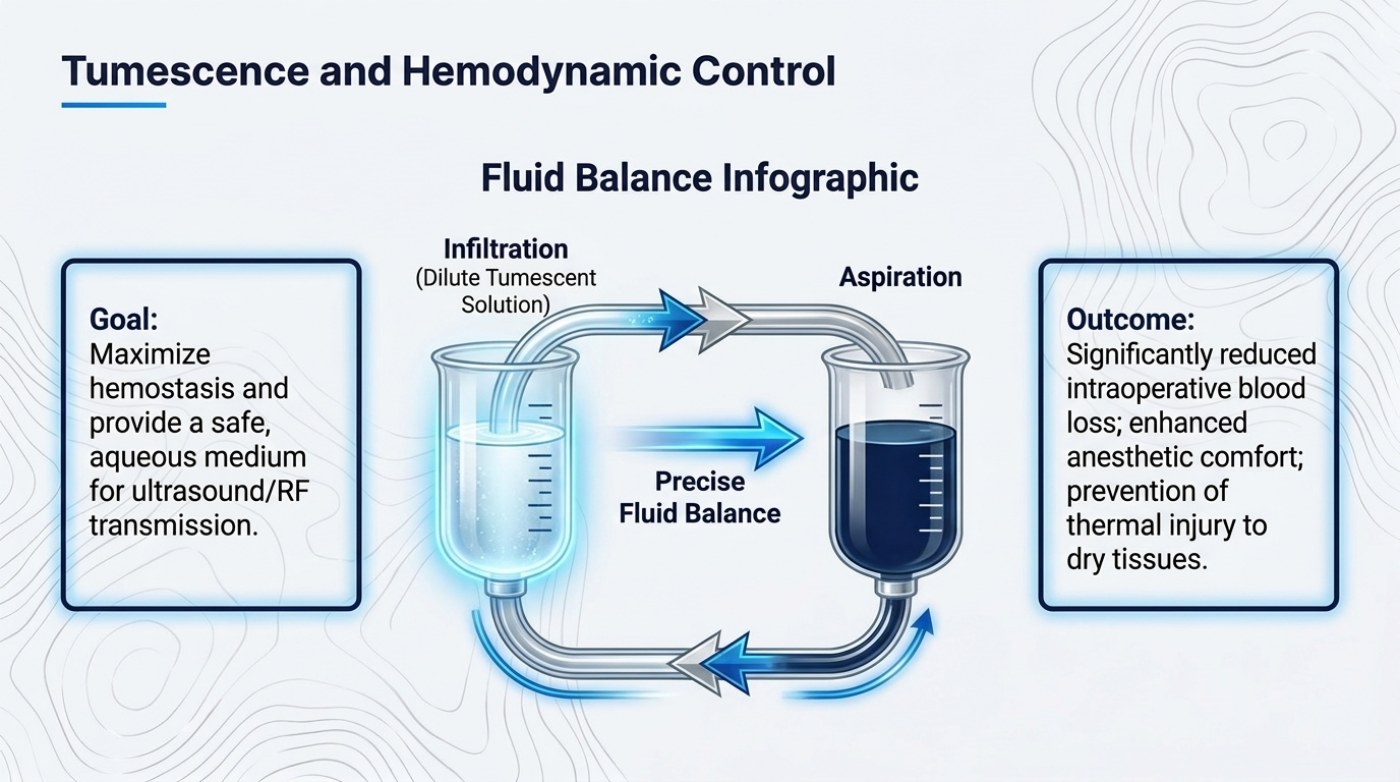
The fluid pathway, stage by stage
The infographic is arranged left to right. On the left, "Infiltration" is glossed in brackets as "Dilute Tumescent Solution" — the fluid that goes in before anything is removed. In the middle, an arrow labelled "Precise Fluid Balance" connects the two sides. On the right, "Aspiration" is the stage at which fat and fluid come out. The two text blocks then frame the sequence: the goal describes what infiltration is for, and the outcome describes what the whole pathway is supposed to achieve.
What makes the diagram worth reading carefully is that the middle stage is drawn as its own step rather than as a by-product. Fluid balance is presented as something tracked, not something assumed. That framing matters because the volume of fluid involved is not incidental to a large case — the site's published risk guide records the overall complication rate rising from 1.1% to 3.7% above the five-litre aspirate line, with seroma the most common complication driving that step, which makes what goes in as relevant as what comes out.
| Stage on the slide | What happens | What it is for |
|---|---|---|
| Infiltration — dilute tumescent solution | Fluid is placed into the tissue before any fat is removed | Creates the plane and the medium the rest of the operation depends on |
| Precise fluid balance | What goes in is tracked against what comes out | Keeps the balance a measured decision rather than an estimate |
| Aspiration | Fat and fluid are removed through the instruments | Delivers the change in contour |
| Stated goal | Maximise hemostasis and provide a safe, aqueous medium for ultrasound/RF transmission | Less bleeding during the case, and a medium the energy devices can work through |
| Stated outcome | Significantly reduced intraoperative blood loss; enhanced anesthetic comfort; prevention of thermal injury to dry tissues | Names the three effects the slide groups as the point of the exercise |
What the dilute solution is doing in the tissue
The slide's goal block names two functions and the outcome block names three effects, and the pairing is worth separating because they act on different things. Hemostasis is a vascular effect: the solution is described as maximising it, which means the field bleeds less while work is happening. The aqueous medium is a physical effect: it is described as making the tissue safe for ultrasound and radiofrequency transmission, which is what allows the energy methods from the deck's earlier slides to be used at all.
The outcome block then lists what those functions are meant to deliver. Significantly reduced intraoperative blood loss is the hemostasis effect made visible. Enhanced anesthetic comfort is a separate benefit of the same infiltration step, described by the slide as comfort during and after the case rather than as a named agent or amount. Prevention of thermal injury to dry tissues is the aqueous medium's contribution to safety: the site's published tightening guide covers the thermal side of this in more detail, pairing a 65 °C subdermal working target with a 42 °C surface cap, and notes the operating-room environment — an ambient range of 20–23 °C with positive-pressure ventilation — that supports heat dissipation. The slide's point is narrower and simpler: tissue that contains fluid behaves differently from tissue that does not.
| Function | Mechanism as the slide describes it | Why it matters to the result |
|---|---|---|
| Hemostasis | Prepared infiltration maximises hemostasis | Bleeding at the moment of removal is what produces bruising and obscures the working plane |
| Aqueous medium | Provides a safe medium for ultrasound and radiofrequency transmission | Energy devices behave differently in wet tissue than in dry tissue |
| Anesthetic comfort | A dilute solution carries local anaesthetic into the area | Described as comfort during and after the case, not as a named agent or amount |
| Thermal protection | The slide names prevention of thermal injury to dry tissues as an outcome | The same heating that serves contraction can burn skin when it is not controlled |
| Separation of layers | Fluid enters the tissue before any fat is removed | Makes layer-specific work possible instead of removing fat indiscriminately |
Two beakers, one system
The drawing's left beaker is pale and being filled; the right beaker is dark and being drained; the two are joined by a path that curves underneath them and doubles back, with arrows running both ways. The visual claim is that infiltration and aspiration are not two separate events with a gap between them. They are one circuit, and the fluid that goes in is largely what comes back out, mixed with what was removed.
That is why the slide puts blood loss in the outcome block rather than in a complications list. In a case run on this principle, the fluid that leaves the body is mostly the fluid that was put in, which is a very different situation from an operation in which the tissue is cut in its natural state. The site's published figures support the direction of travel: in a prospective study of 56 patients who received equal volumes of traditional and laser-assisted liposuction at two contralateral sites, blood loss fell by more than 50% on the laser-treated side, and published comparisons of suction-assisted against ultrasound-assisted techniques measure the same category of difference.
Why the middle arrow is the control point
"Precise Fluid Balance" is printed in the centre of the diagram, between the beakers, and it is the only stage described as precise. That word choice is the slide's clinical argument. Fluid that is infiltrated and not accounted for, or aspirated beyond what was planned, is how a case moves from a controlled procedure into a fluid problem — and fluid is where the published complication data concentrates. The site's published risk guide notes that the ASPS TOPS analysis of more than 4,500 patients found seroma the most common complication overall, and the reason the overall complication rate stepped up above the five-litre aspirate line. The site's etching guide records a 50-patient drain study in which 17 patients developed a post-operative seroma — 13 of 25 where no adjunctive drainage was used against 4 of 25 where drainage was added — with volumes graded as mild under 50 cc, moderate 50–100 cc and severe over 100 cc.
None of those figures belong to the slide, and none of them predicts an individual course. What they do is show why the diagram puts the balance step in the middle: it is the place where the operation's fluid arithmetic is either kept inside known limits or allowed to drift.
| Published anchor | Figure | Already carried in |
|---|---|---|
| Blood loss with an energy-assisted technique | More than 50% reduction across 56 patients treated at two contralateral sites | The site's published risk guide |
| Seroma with and without adjunctive drainage | 17 of 50 patients overall; 13 of 25 without drainage versus 4 of 25 with it | The site's etching and risk guides |
| Seroma severity scale | Mild under 50 cc, moderate 50–100 cc, severe over 100 cc | The site's etching guide |
| Volume threshold for the whole procedure | Overall complication rate rises from 1.1% to 3.7% above five litres of aspirate | The site's published risk guide |
Frequently asked questions
Why infiltrate fluid before removing fat at all?
The slide gives two reasons in its goal block: to maximise hemostasis, so the field bleeds less, and to provide a safe aqueous medium for ultrasound and radiofrequency transmission. A third follows from the first — reducing bleeding keeps the working plane visible, which is what makes layer-specific removal possible rather than bulk removal.
What does "precise fluid balance" mean in practice?
It means the fluid put in is tracked against the fluid taken out, so the volume decision stays a measured one. The slide draws it as its own stage between infiltration and aspiration rather than as an incidental by-product, and the published data already summarised on this site explain why fluid is where the complication rate concentrates as case volume rises.
Does tumescent infiltration remove the risk of bleeding or of fluid complications?
No. It changes the operating conditions and is described as significantly reducing intraoperative blood loss, but published rates for seroma and other fluid complications remain, and they step up with volume — the overall complication rate rising from 1.1% to 3.7% above five litres of aspirate in the analysis carried on this site. Risk reduction is not risk removal, and individual risk is a question for an examining surgeon.
Related reading
- Liposuction Risks and Complication Rates as Published
- How Much Fat Can Be Removed in One Liposuction Session?
- Liposuction Recovery Week by Week: Compression, Massage and Exercise Milestones
This is published information, not medical advice — a board-certified surgeon must assess whether a procedure suits you.